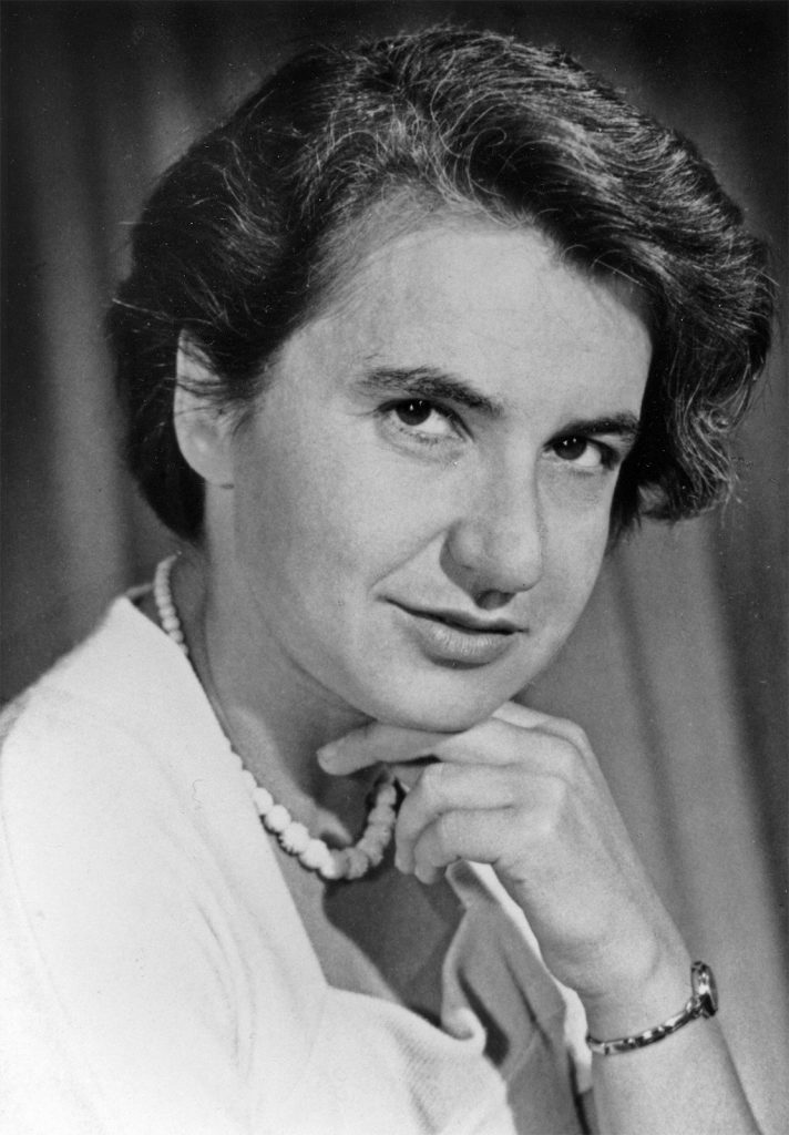
Rosalind Elsie Franklin was born in London, England July 25, 1920 and despite a tragically short life (she died in April 1958 from ovarian cancer) she made tremendous scientific achievements in figuring out what molecules look like and how that relates to what they do. Best known for “Photo 51,” the “blurry X” of an x-ray diffraction pattern showing that DNA forms a specific double helix, Franklin also was a pioneer of carbon technology and she discovered innerworkings of viruses including polio and tobacco mosaic virus (TMV).
Rosalind’s father wanted to become a scientist but WWI got in the way – and he almost got in the way of Franklin becoming a scientist. He discouraged Rosalind from pursuing her goal of becoming a scientist (which she had her hearts set on by the age of 15) – not out of malice, but because it was a very difficult career path for a woman. But, while Rosalind couldn’t be dissuaded, her father was able to be convinced. So, after attending one of the only schools at the time to teach girls physics and chemistry, St. Paul’s Girls’ School, she enrolled at Cambridge University’s Newnham College in 1938 as a chemistry student.
After graduating in 1941, she received a scholarship to carry out graduate research. She used the scholarship to spend a year in photochemist R.G.W Norrish’s lab, then took a position as an assistant research officer for the British Coal Utilization Research Association (CURA). She worked there until 1947, studying the physical structure of coal – and publishing numerous papers on the topic that are still widely-cited. Among other things, she found out that different forms of carbon can form different “meshes” at the molecular level that can filter out and trap various other molecules. By determining that different types of coal have different microstructures – and that their porosity is temperature-dependent – she was able to classify different types of coal and predict their usefulness for different tasks. Her work also helped make possible carbon fiber technology and earned her a PhD in physical chemistry from Cambridge in 1945.
After CURA, she then moved to Paris, where an old friend helped her get in touch with Marcel Mathieu, a big-wig in French research at the time. Mathieu saw the potential for Franklin to be great too and offered her a position as a “chercheur” in the Laboratoire Central des Services Chimiques de l’Etat. It was here where, through the tutelage of Jacque Mering, she learned X-ray diffraction techniques (which would later make her not-famous-enough). X-ray diffraction techniques beam x-rays at molecular samples – the x-rays scatter when they hit the sample and scientists can then use the scattered x-rays to figure out where they scattered from.
She took this newfound technical prowess back to England where, in 1951, a Turner and Newall Fellowship in hand, she went to work as a research associate at King’s College London in John Randall’s Biophysics Unit. Randall assigned Franklin a project to work on DNA structural studies while another researcher, Maurice Wilkins was away. When Wilkins came back, he acted as if Franklin were merely his technical assistant instead of being her own research group leader. After getting off on the wrong foot, things didn’t improve in their relationship (not helped by the fact that women at the time couldn’t even eat lunch in the same room as the men did).
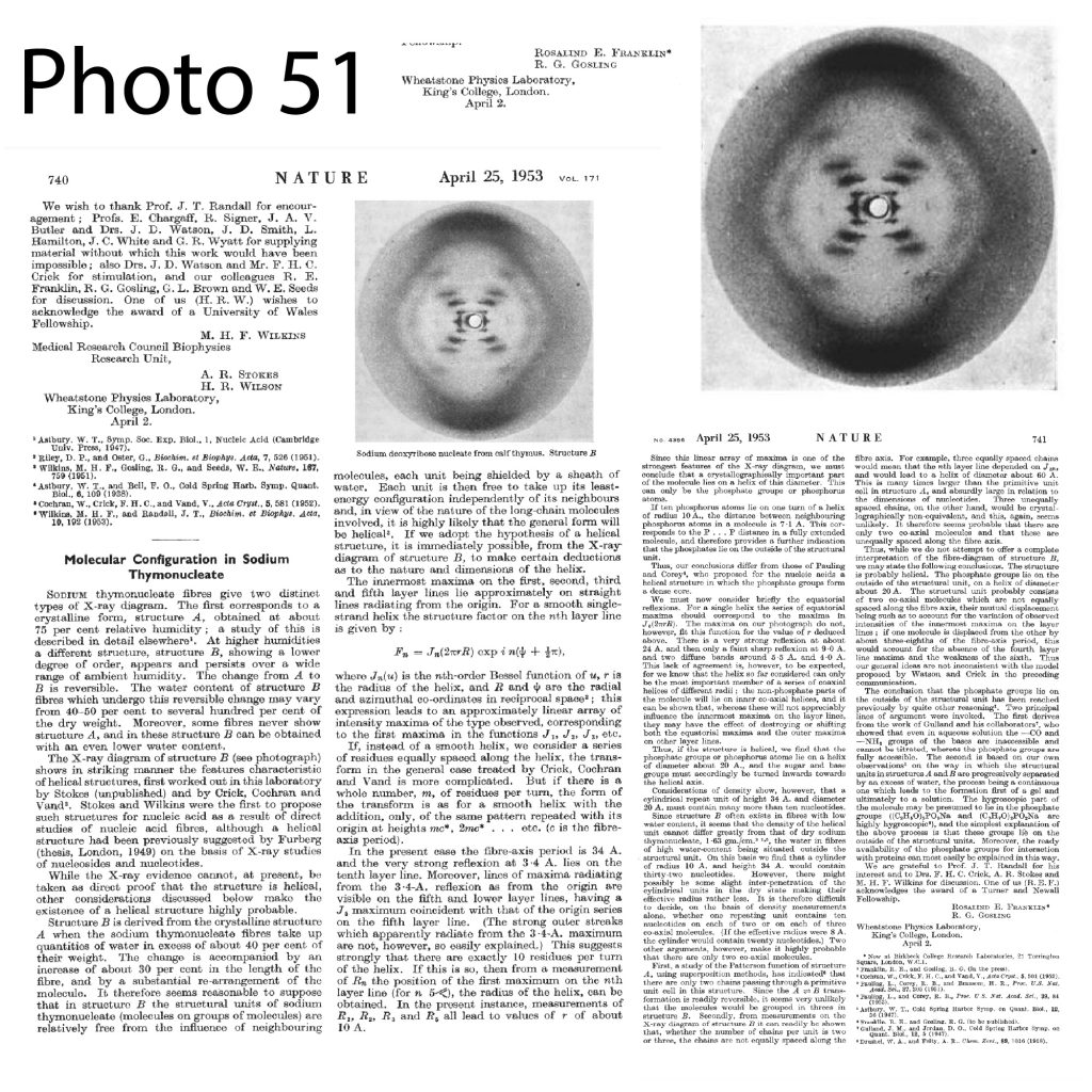
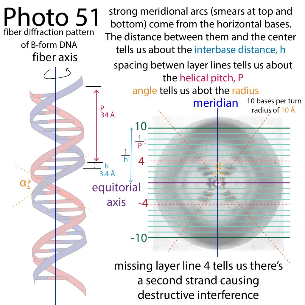
Despite the difficult environment for a female researcher, Franklin was able to made incredible progress in her work. Together with graduate student Raymond Gossling, she figured out how to get informative x-ray diffraction information from DNA. Scientists had been stuck in part because they were only trying to look at a semi-crystal form of DNA, in which the DNA took on an “unnatural” shape and gave a confusing signal. But, by keeping the DNA “wet” and looking at its structure in the soluble fiber form (fiber diffraction instead of crystallography), she was able to capture a “picture” of DNA in its natural form.
This picture held the key information to calculating the geometry of the double helix DNA forms – not just the fact that it’s a double helix with the phosphate groups on the outside and the bases sticking in – but everything from the number of bases per turn, to the vertical spread and the radius. More on how here: http://bit.ly/fiberdiffractiondna
Franklin was on the cusp of unlocking that information when, without her knowledge, let alone approval, Maurice Wilkins showed some of Franklin’s work, including the iconic Photo 51, to Watson and Crick, who were independently carrying out theoretical modeling to figure out DNA structure at Cambridge. The picture provided the missing clues to getting their model to fit, and they were able to speedily publish their classic paper in Nature in 1953 (in a journal issue where Franklin’s Photo 51 paper also appeared but was greatly overshadowed).
Looking for a research environment where she had more autonomy and faced less hostility, Franklin left in 1953 to start her own research group at Birkbeck College in London. As a condition of leaving, Franklin had promised Kings College not to work on DNA, so she switched to the field of virology, studying the structures of viruses including the plant virus tobacco mosaic virus (TMV) and polio, and publishing 17 papers over the next five years (as well as creating giant 3D models fort the 1958 Brussels World’s Fair).
We will never know what other great accomplishments Franklin would have undoubtedly made were her life not cut tragically short by ovarian cancer. She passed away in 1959 at the age of 37 and Watson, Crick, & Wilkins got the Nobel Prize in 1962 for the DNA structure discovery she played a key role in.